Thông tin nghiên cứu
Loại tài liệu
Bài báo trên tạp chí khoa học (Journal Article)
Tiêu đề
Nghiên cứu một số hình ảnh bất thường ở vùng mũi - xoang trên phim CT Scan
Tác giả
Phan Mộng Hoàng; Nghiêm Đức Thuận
Năm xuất bản
2012
Số tạp chí
1
Trang bắt đầu
1-2
ISSN
1859-1868
Nguồn
Từ khóa nghiên cứu
Abstract
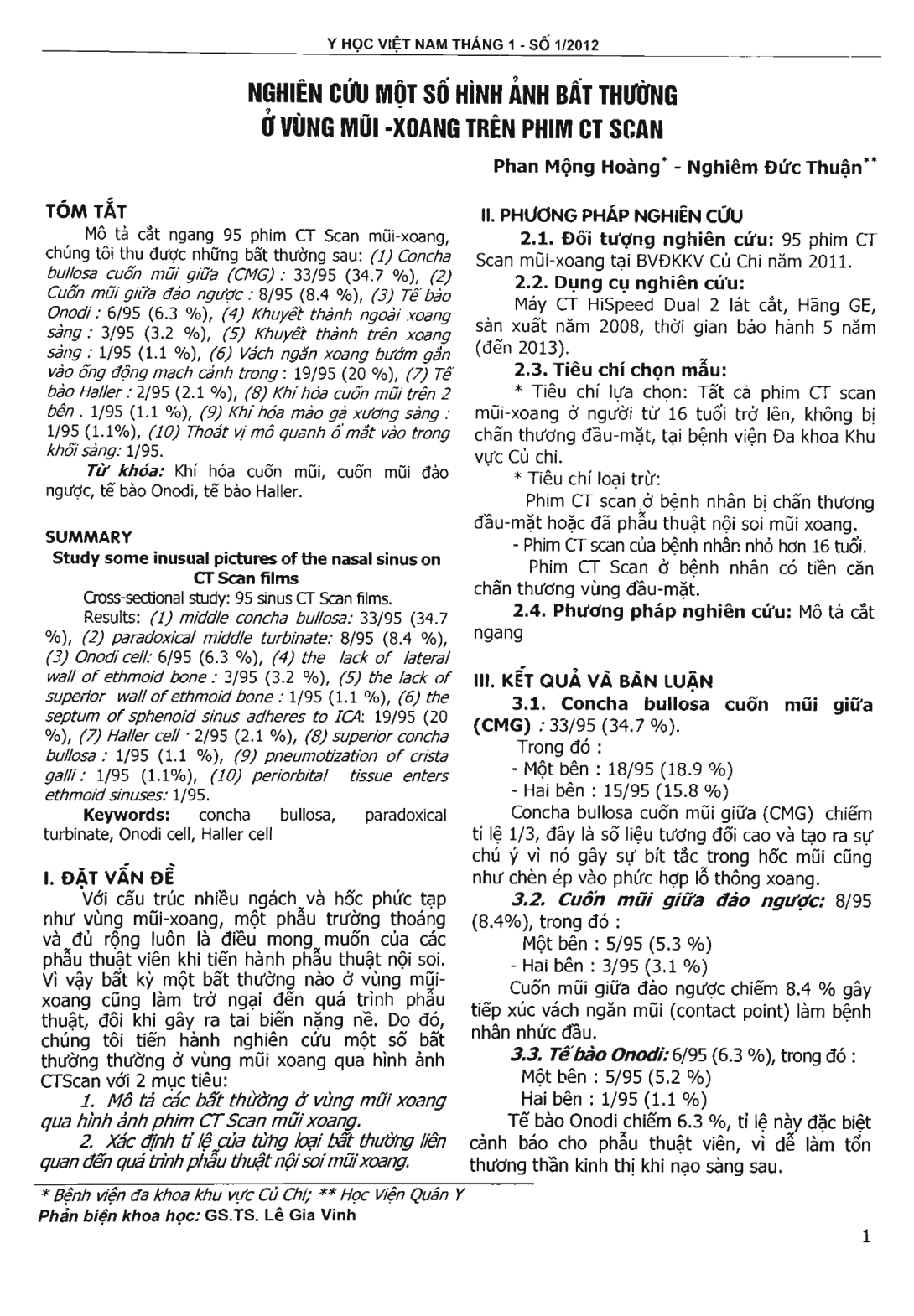
Cross-sectional study: 95 sinus CT Scan films. Results: (1) middle concha bullosa: 33/95 (34.7 percent), (2) paradoxical middle turbinate: 8/95 (8.4 percent), (3) Onodi cell: 6/95 (6.3 percent), (4) the lack of lateral wall of ethmoid bone: 3/95 (3.2 percent), (5) the lack of superior wall of ethmoid bone: 1/95 (1.1 percent), (6) the septum of sphenoid sinus adheres to ICA: 19/95 (20 percent), (7) Haller cell: 2/95 (2.1 percent), (8) superior concha bullosa: 1/95 (1.1 percent), (9) pneumotization of crista galli: 1/95 (1.1 percent), (10) periorbital tissue enters ethmoid sinuses: 1/95.
- Đăng nhập để gửi ý kiến
